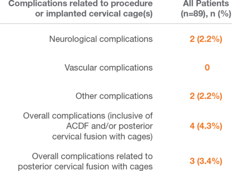
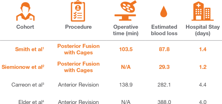
Clinical Evidence | Providence

Low Rate of Complications*

  • A retrospective, multicenter review of prospectively collected data performed at 11 centers.
  • Follow‑up evaluation included clinical status and pain scale (VAS – visual analog scale).
  • The procedure has a favorable overall complication profile, a short length of hospital stay (29 hours), and negligible blood loss.*
  • The overall complication rate for posterior cervical cages was 3.4%.
  • This complication rate compares favorably to the 12.33% overall complication profile reported for ACDF/arthroplasty.1–13
  • Posterior cervical fusion with cages can be considered a safe alternative for patients undergoing cervical spine surgery.
* Compared to ACDF/arthroplasty, corpectomy, laminectomy, laminoplasty

References

  1. Q, Bi X, Ni B, Lu X, Chen J, Yang J, et al. Outcomes of three anterior decompression and fusion techniques in the treatment of three‑level cervical spondylosis. Eur Spine J 2011;20:1539‑44.
  2. Lian XF, Xu JG, Zeng BF, Zhou W, Kong WQ, Hou TS. Noncontiguous anterior decompression and fusion for multilevel cervical spondylotic myelopathy: A prospective randomized control clinical study. Eur Spine J 2010;19:713‑9.
  3. Uribe JS, Sangala JR, Duckworth EA, Vale FL. Comparison between anterior cervical discectomy fusion and cervical corpectomy fusion using titanium cages for reconstruction: Analysis of outcome and long‑term follow‑up. Eur Spine J 2009;18:654‑62.
  4. Nirala AP, Husain M, Vatsal DK. A retrospective study of multiple interbody grafting and long segment strut grafting following multilevel anterior cervical decompression. Br J Neurosurg 2004;18:227‑32.
  5. Song KJ, Lee KB, Song JH. Efficacy of multilevel anterior cervical discectomy and fusion versus corpectomy and fusion for multilevel cervical spondylotic myelopathy: A minimum 5‑year follow‑up study. Eur Spine J 2012;21:1551‑7. 20. Liu Y, Hou Y, Yang L, Chen H, Wang X, Wu X, et al. Comparison of 3 reconstructive techniques in the surgical management of multilevel cervical spondylotic myelopathy. Spine (Phila Pa 1976) 2012;37:E1450‑8.
  6. Lin Q, Zhou X, Wang X, Cao P, Tsai N, Yuan W. A comparison of anterior cervical discectomy and corpectomy in patients with multilevel cervical spondylotic myelopathy. Eur Spine J 2012;21:474‑81.
  7. Hwang SL, Lee KS, Su YF, Kuo TH, Lieu AS, Lin CL, et al. Anterior corpectomy with iliac bone fusion or discectomy with interbody titanium cage fusion for multilevel cervical degenerated disc disease. J Spinal Disord Tech 2007;20:565‑70.
  8. Yonenobu K, Fuji T, Ono K, Okada K, Yamamoto T, Harada N. Choice of surgical treatment for multisegmental cervical spondylotic myelopathy. Spine (Phila Pa 1976) 1985;10:710‑6.
  9. Bertalanffy H, Eggert HR. Complications of anterior cervical discectomy without fusion in 450 consecutive patients. Acta Neurochir (Wien) 1989;99:41‑50.
  10. Burke JP, Gerszten PC, Welch WC. Iatrogenic vertebral artery injury during anterior cervical spine surgery. Spine J 2005;5:508‑14.
  11. Emery SE, Bohlman HH, Bolesta MJ, Jones PK. Anterior cervical decompression and arthrodesis for the treatment of cervical spondylotic myelopathy. Two to seventeen‑year follow‑up. J Bone Joint Surg Am 1998;80:941‑51. Journal of Craniovertebral Junction and Spine / Volume 8 / Issue 4 / October-December 2017 349
  12. Jamjoom ZA. Pharyngo‑cutaneous fistula following anterior cervical fusion. Br J Neurosurg 1997;11:69‑74.

Independent Prospective Randomized Study

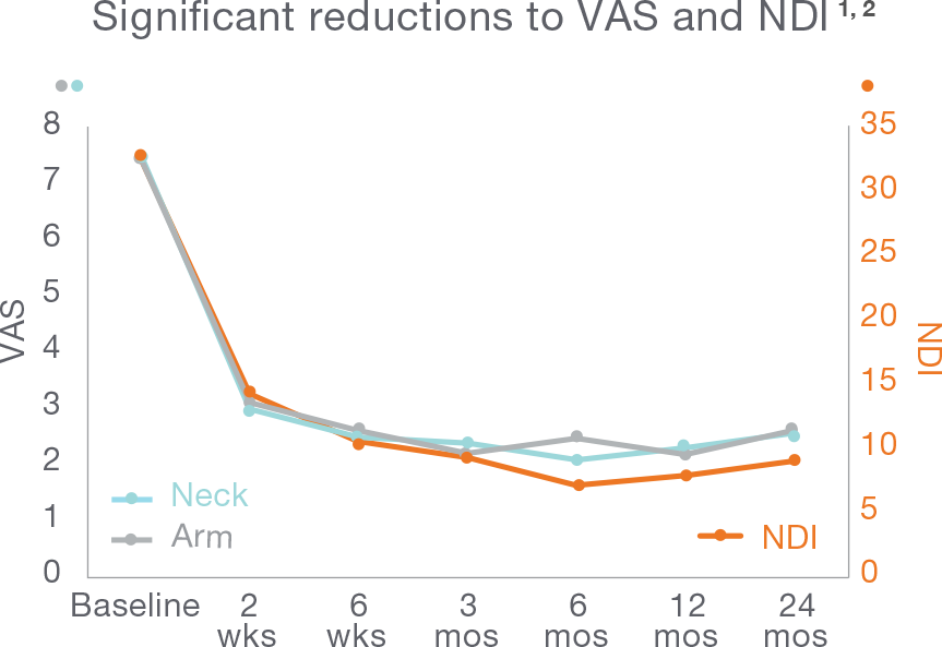
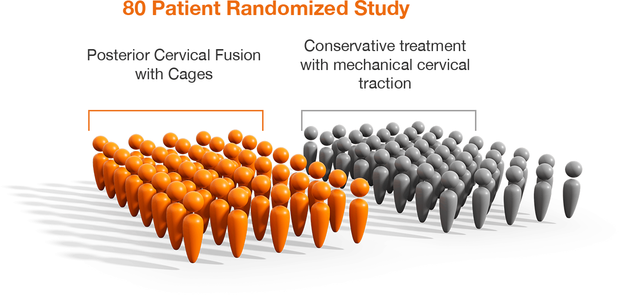
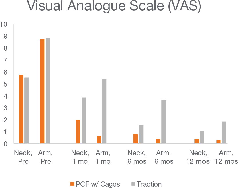
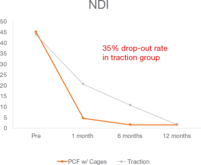
PCF with Cages vs. Traction Single Level Cervical Radiculopathy 80 Patients @ 1-year follow-up
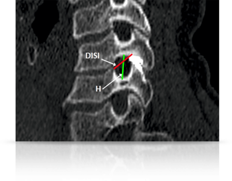
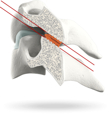
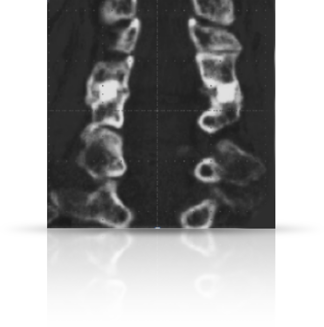
  • 80 patients randomized into two groups—a surgical group in which patients were given posterior cervical fusion with cages and a traction group in which patients were treated conservatively with mechanical cervical tractions.
  • Visual analog scale for arm and neck, Neck Disability Index, and—Short Form-36 (SF-36) questionnaires administered preoperatively and after treatment up to 12 months.
  • Posterior cervical fusion with cages is a safe and effective procedure to treat single-level cervical radiculopathy.

Foraminal Expansion & Nerve Root Decompression

  • Radiographic analysis of 43 subjects enrolled in a prospective, multi-center study.
  • CT scans were obtained at baseline and 6- and 12-months after cervical fusion using bilateral posterior cervical cages.
  • Cervical cages placed bilaterally in the facet joints from a posterior approach significantly increase foraminal area.
  • Foraminal height, area, theoretical area, and inferior diagonal without implant (DISI) were significantly greater following implant placement.
Team | Innovation | Quality | Passion | Transparency
Copyright © 2023, Providence Medical Technology. All rights reserved.