ENABLING TOTAL SEGMENTAL FUSION. ENGINEERED FOR OSSEOINTEGRATION.

New porous titanium architecture.
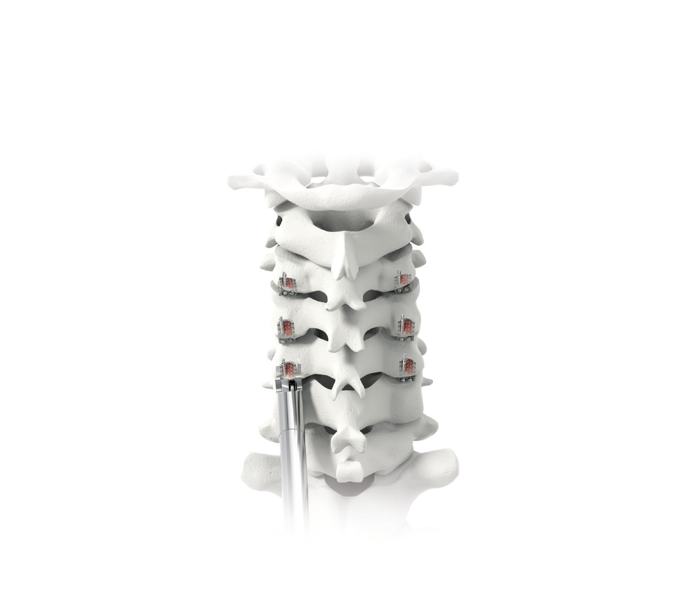

The Next Dimension in Posterior Fixation

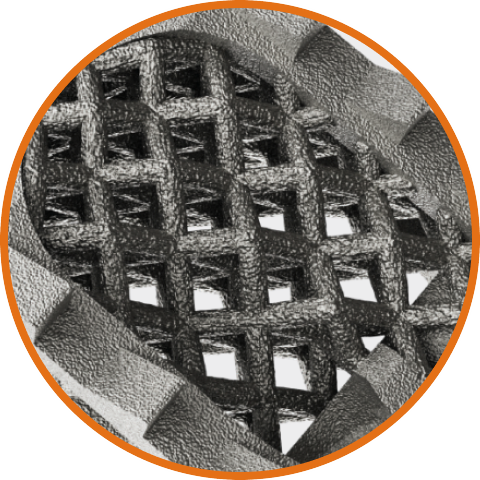
CORUS™ PCSS 3D–T features a pore structure consistent with those shown in published literature to promote vascularization and osseointegration.¹⁻⁴
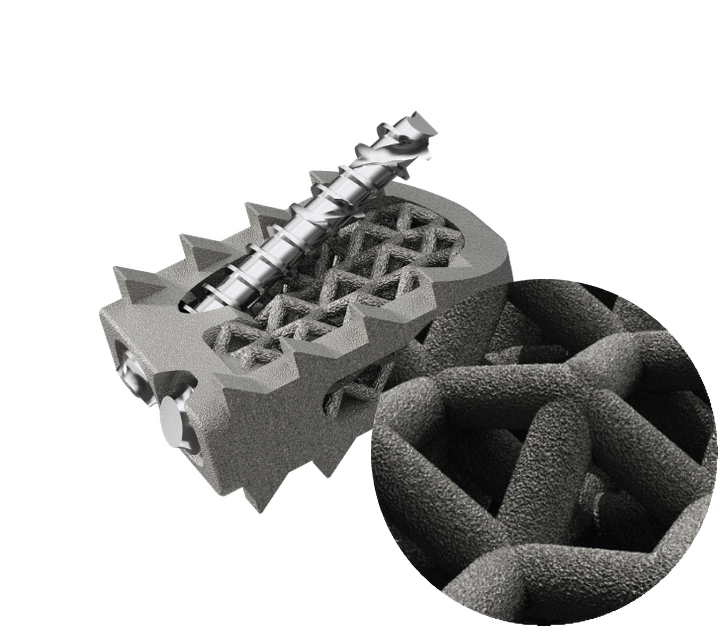

SIMILAR TO CANCELLOUS BONE ⁵˒⁶˒⁷

Interconnected lattice provides a scaffold for bony ingrowth
Overall porosity 73–75%
Pore size range ~400–800 μm
Average pore size 609 μm

INCREASED SURFACE AREA AND ROUGHNESS

1.5X greater surface area vs machined PCSS⁸
8X surface roughness vs machined PCSS⁹

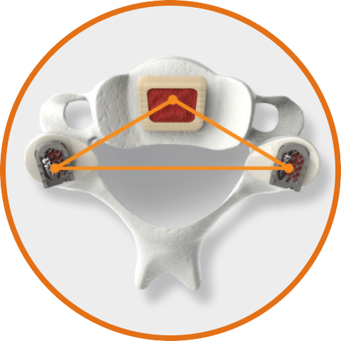
Supports Multilevel Fusion

Circumferential cervical fusion is associated with greater biomechanical stability and higher fusion rates in multilevel cervical fusion. Tissue-sparing posterior techniques allow surgeons to add supplemental posterior fixation and fusion without increasing hospital length of stay.¹⁰

Proprietary Porous Lattice

Designed for isotropic load distribution (absorbing force equally from all directions) while providing open pathways for vascularization and bony ingrowth.

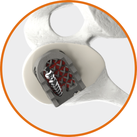
Enhanced Anatomical Fit

Optimized geometry to address variable patient anatomy.

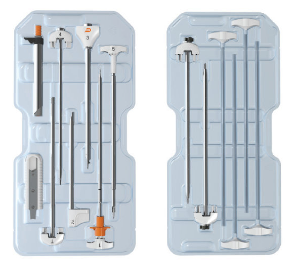
Designed for Perioperative Efficiency

  • Sterile-packaged & single-use
  • Numbered instruments
  • Tissue-sparing technique*
* CORUS PCSS 3D-T supports tissue-sparing portal-based, mini-open, fluoroscopic, and navigated techniques as well as open procedures based on surgeon preference and patient anatomy.

As with all medical devices, there are risks and considerations to device use. Please refer to the device labeling for a full discussion of potential risks, contraindications, warnings, precautions, and instructions for use. Rx only.

Treatment may not be appropriate for all patients and all patients may not benefit. Results vary.

Indications for Use

Indications for Use, CORUS™ PCSS 3D-T

CORUS™ Posterior Cervical Stabilization System 3D-T (CORUS PCSS 3D-T) is posterior spinal instrumentation with integrated screw fixation intended to provide immobilization and stabilization of spinal segments. CORUS PCSS 3D-T is placed through a posterior surgical approach in up to 3 consecutive levels of the cervical spine (C3–C7) and achieves bilateral facet fixation by spanning the facet interspace at each level with points of fixation at each end of the construct.

CORUS PCSS 3D-T is intended as an adjunct to posterior cervical fusion (PCF) and is only intended to be used in combination with an FDA cleared/approved spine stabilization hardware for anterior cervical fusion at the same level(s).

CORUS PCSS 3D-T is indicated for skeletally mature patients with degenerative disc disease (DDD). DDD is defined as radiculopathy and/or myelopathy, neck and/or arm pain of discogenic origin as confirmed by radiographic studies. CORUS PCSS 3D is to be used with autogenous bone and/or allogenic bone graft.

Indications for Use, CORUS™ Spinal System

CORUS™ Spinal System is a set of instruments indicated to be used to perform posterior cervical fusion in patients with cervical degenerative disc disease.


View Safety Information MKT-PMT-861 CORUS 3D-T Webpage

References

  1. Taniguchi N, et al. Effect of pore size on bone ingrowth into porous titanium implants fabricated by additive manufacturing: An in vivo experiment. Mater Sci Eng C Mater Biol Appl. 2016 Feb;59:690-701. doi:10.1016/j.msec.2015.10.069. PMID: 26652423.
  2. Ran Q, Yang W, Hu Y, Shen X, Yu Y, Xiang Y, Cai K. Osteogenesis of 3D printed porous Ti6Al4V implants with different pore sizes. J Mech Behav Biomed Mater. 2018 Aug;84:1-11. doi:10.1016/j.jmbbm.2018.04.010. PMID: 29709846.
  3. McGilvray KC, Easley J, Seim HB, Regan D, Berven SH, Hsu WK, Mroz TE, Puttlitz CM. Bony ingrowth potential of 3D-printed porous titanium alloy: a direct comparison of interbody cage materials in an in vivo ovine lumbar fusion model. Spine J. 2018 Jul;18(7):1250-1260. doi:10.1016/j.spinee.2018.02.018. PMID: 29496624.
  4. Egan PF, Gonella VC, Engensperger M, Ferguson SJ, Shea K. Computationally designed lattices with tuned properties for tissue engineering using 3D printing. PLoS One. 2017 Aug 10;12(8):e0182902. doi:10.1371/journal.pone.0182902. PMID: 28797066.
  5. RPT-PMT-591 Rev 0, Engineering Report Providence Medical Lattice Characterization.
  6. Keaveny TM, Morgan EF, Niebur GL, Yeh OC. Biomechanics of Trabecular Bone. Annu Rev Biomed Eng. 2001;3:307-33.
  7. Karageorgiou V, Kaplan D. Porosity of 3D Biomaterial Scaffolds and Osteogenesis. Biomaterials. 2005 Sep;26(27):5474-91.
  8. RPT-PMT-597 Surface Area Analysis Report, Cervical Cage-X and 3D Stabilizer.
  9. RPT-PMT-592 Surface Roughness Characterization Report, Covalent Metrology.
  10. Strenge KB, et al. Three-Level Anterior Cervical Discectomy and Fusion With or Without an Investigational Posterior Stabilization System Assessed Through 24 Months: A Multicenter Randomized Controlled Trial. Spine (Phila Pa 1976). 2025 Sep 1.
Team | Innovation | Quality | Passion | Transparency
Copyright © 2025, Providence Medical Technology. All rights reserved.

Our Mission is to establish Circumferential Cervical Fusion (CCF) as the standard of care for high-risk patients.


Copyright © 2026, Providence Medical Technology. All rights reserved.